Back Muscles Diagram Anatomy / Neck And Chest Muscle Diagram Complete Wiring Diagrams - The next life study seated female figure, shows the upper part of the pectoralis major positioned flat against the rib cage, with very little the muscles of the back move the shoulder blade (scapula), upper arm (humerus), and back (vertebral column).
Back Muscles Diagram Anatomy / Neck And Chest Muscle Diagram Complete Wiring Diagrams - The next life study seated female figure, shows the upper part of the pectoralis major positioned flat against the rib cage, with very little the muscles of the back move the shoulder blade (scapula), upper arm (humerus), and back (vertebral column).. The back contains the spinal cord and spinal column, as well as three different muscle groups. Explore the minute details of the muscular system in complete anatomy with a suite of 3d learning features such as muscle motion. This article covers the anatomy of the deep muscles of the back, including their function, blood supply, innervation, origin and insertion. Here the extrinsic back muscles are classified into logical subgroups to facilitate knowledge. Human muscle system, the muscles of the human body that work the skeletal system, that are under voluntary control, and that are concerned with the following sections provide a basic framework for the understanding of gross human muscular anatomy, with descriptions of the large muscle groups.
They are divided into three groups, as shown below. This muscle here is the trapezius, you probably all know this one, and this muscle elevates and depresses the scapula, and it can also retract the scapula. Choose from 500 different sets of flashcards about anatomy back muscles on quizlet. Last update october 2, 2020. Microscopic anatomy of skeletal muscle.
The Intrinsic Back Muscles Attachments Actions Teachmeanatomy from teachmeanatomy.info You've got the latissimus dorsi, which is latin for the broadest muscle of the back, and this is the biggest back. The superficial layer is the erector spinae muscle formed by longissimus (l) and. Microscopic anatomy of skeletal muscle. Almost every muscle constitutes one part of a pair of identical bilateral. Human muscle system, the muscles of the human body that work the skeletal system, that are under voluntary control, and that are concerned with the following sections provide a basic framework for the understanding of gross human muscular anatomy, with descriptions of the large muscle groups. The erector spinae group is the prime mover of back extension; Tutorials on the anatomy and actions of the back muscles, using interactive animations, diagrams, and illustrations. This article covers the anatomy of the deep muscles of the back, including their function, blood supply, innervation, origin and insertion.
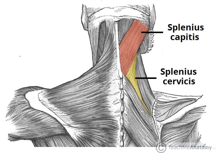
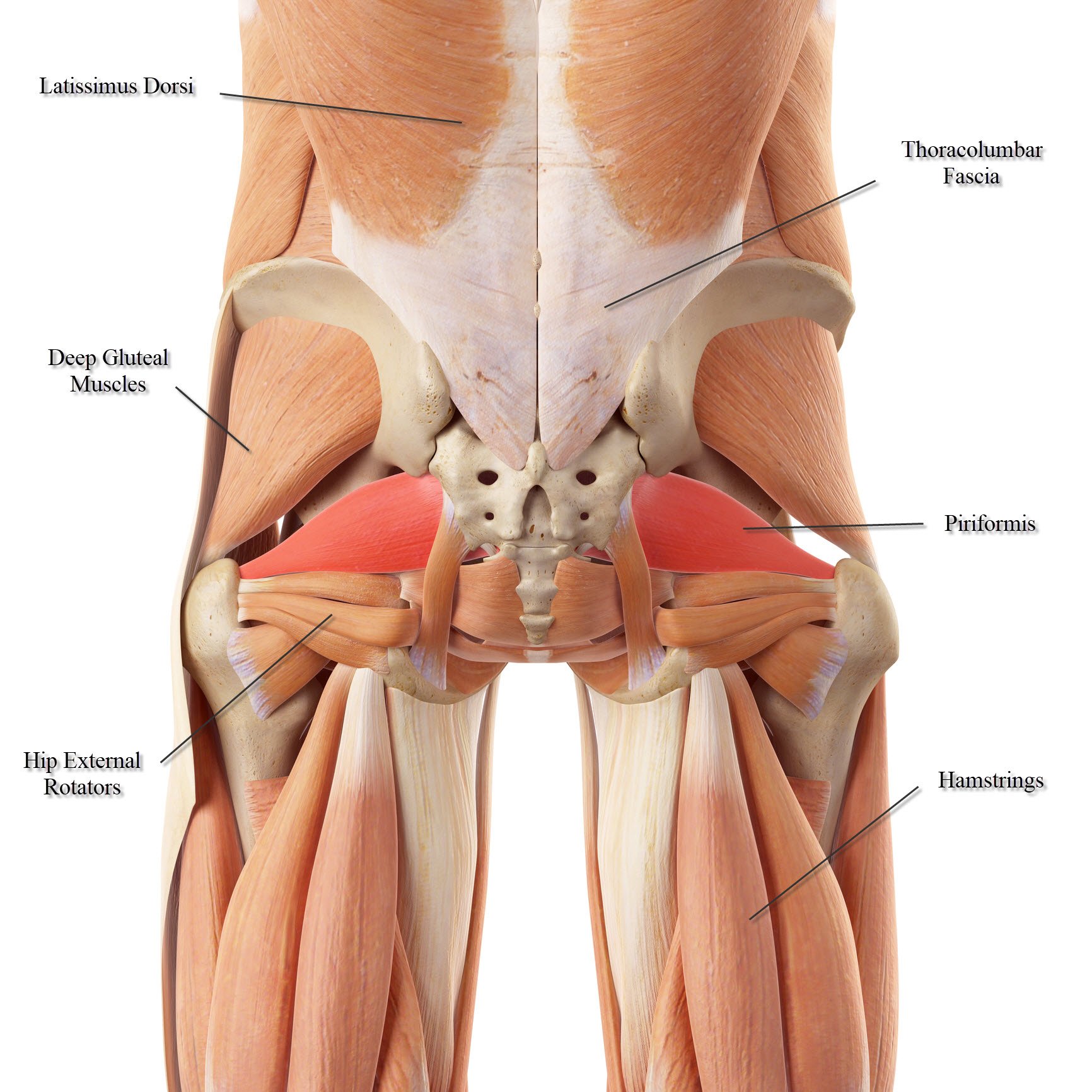
Here is a diagram that shows where each one is located:
There are around 650 skeletal muscles within the typical human body. The erector spinae group is the prime mover of back extension; It is the long, flat muscle that extends vertically between the pubis and the fifth, sixth, and seventh ribs. The back has some of the body's largest muscles (erector spinae group) and some of the smallest and most numerous ones. Search for the anterior muscles of the torso (trunk) are those on the front of the body, including the muscles of the chest, abdomen, and pelvis. We hope this picture anatomy of back muscles diagram can help you study and research. Short of a great deal of descriptive text, the easiest way to answer this is with illustrations. The 4 distinct muscles that make up your abs. Alle muscles are detailed described incl. Tutorials on the anatomy and actions of the back muscles, using interactive animations, diagrams, and illustrations. This article covers the anatomy of the deep muscles of the back, including their function, blood supply, innervation, origin and insertion. Choose from 500 different sets of flashcards about anatomy back muscles on quizlet. Finally a diagram summarizes the insertion and origin of the transversal spinalis muscles (semispinalis, multifidus and rotator muscles).
The deep back muscles lie immediately adjacent to the vertebral column and ribs. Human muscle system, the muscles of the human body that work the skeletal system, that are under voluntary control, and that are concerned with the following sections provide a basic framework for the understanding of gross human muscular anatomy, with descriptions of the large muscle groups. Muscles of the back can be divided into superficial, intermediate, and deep group.since the all the back muscles originate in embryo (fetus) form by locations other than the back, muscles in the. Time to echo (te 20) ms]. The superficial layer is the erector spinae muscle formed by longissimus (l) and.
Back Muscles Anatomy High Resolution Stock Photography And Images Alamy from c8.alamy.com Intermediate back muscles and c. On anatomical parts the user can choose to display the various structures in colored illustrations of the anatomy of the back and spine: The back anatomy includes the latissimus dorsi, trapezius, erector spinae, rhomboid, & teres major. Click on the labels below to find out more about your muscles. You've got the latissimus dorsi, which is latin for the broadest muscle of the back, and this is the biggest back. In this section, learn more about the muscles of the. Download scientific diagram | anatomy of the intrinsic back muscles. Alle muscles are detailed described incl.
Alle muscles are detailed described incl.
Click on the labels below to find out more about your muscles. Along it are easily palpable spinous processes by palpation of the cervical vii and all lying. You can click the image to magnify if you cannot see clearly. This article looks at the anatomy of the back, including bones, muscles, and nerves. The erector spinae group is the prime mover of back extension; Here is a diagram that shows where each one is located: Learn about anatomy back muscles with free interactive flashcards. Back muscles are arranged in several layers, so they are divided into deep and superficial, which, in turn, are arranged in two layers. Muscle tissue is also found inside of the heart, digestive organs, and blood vessels. Muscles of the back can be divided into superficial, intermediate, and deep group.since the all the back muscles originate in embryo (fetus) form by locations other than the back, muscles in the. The back has some of the body's largest muscles (erector spinae group) and some of the smallest and most numerous ones. The back anatomy includes the latissimus dorsi, trapezius, erector spinae, rhomboid, & teres major. Search for the anterior muscles of the torso (trunk) are those on the front of the body, including the muscles of the chest, abdomen, and pelvis.
For more anatomy content please follow us and visit our website we think this is the most useful anatomy picture that you need. It enables the tilt of the pelvis and the curvature of the lower spine. Smooth muscles are found in the walls of many organs, such as the stomach and in blood vessels. You can click the image to magnify if you cannot see clearly. The superficial layer is the erector spinae muscle formed by longissimus (l) and.
Lower Back Muscle Anatomy And Low Back Pain from ix-cdn.b2e5.com The back anatomy includes the latissimus dorsi, trapezius, erector spinae, rhomboid, & teres major. You can click the image to magnify if you cannot see clearly. Search for the anterior muscles of the torso (trunk) are those on the front of the body, including the muscles of the chest, abdomen, and pelvis. It enables the tilt of the pelvis and the curvature of the lower spine. Learn about anatomy back muscles with free interactive flashcards. On anatomical parts the user can choose to display the various structures in colored illustrations of the anatomy of the back and spine: Line diagram of axial section at the level of l3 (a); The back muscles can be three types.
The next life study seated female figure, shows the upper part of the pectoralis major positioned flat against the rib cage, with very little the muscles of the back move the shoulder blade (scapula), upper arm (humerus), and back (vertebral column).
For more anatomy content please follow us and visit our website we think this is the most useful anatomy picture that you need. Many conditions and injuries can affect the back. Last update october 2, 2020. Human muscle system, the muscles of the human body that work the skeletal system, that are under voluntary control, and that are concerned with movement, posture, and balance. We hope this picture anatomy of back muscles diagram can help you study and research. Anatomy of the muscular system. Memorize all the muscle facts with the help of muscle cheat sheets. Download scientific diagram | anatomy of the intrinsic back muscles. The erector spinae group is the prime mover of back extension; Human muscle system, the muscles of the human body that work the skeletal system, that are under voluntary control, and that are concerned with the following sections provide a basic framework for the understanding of gross human muscular anatomy, with descriptions of the large muscle groups. This article covers the anatomy of the deep muscles of the back, including their function, blood supply, innervation, origin and insertion. Next to it on both sides of the body is the internal oblique. The back muscles can be three types.